Gallery
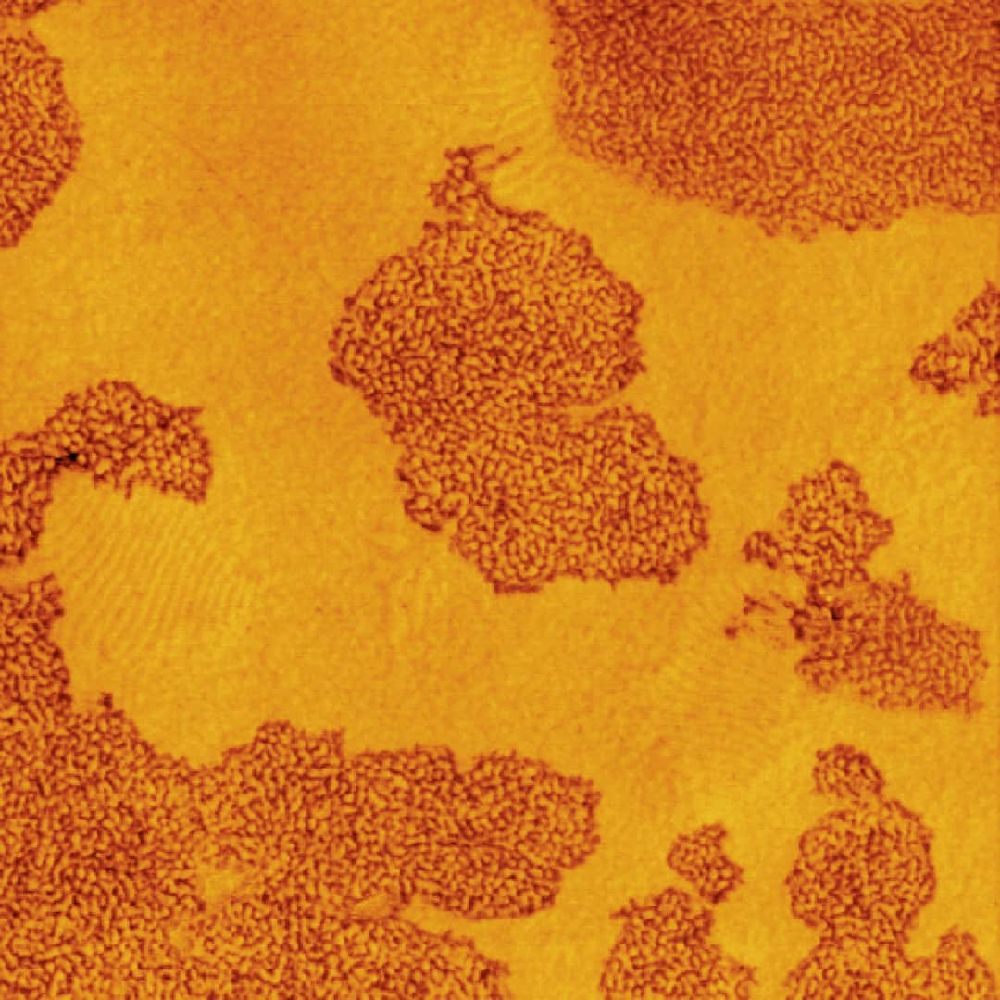
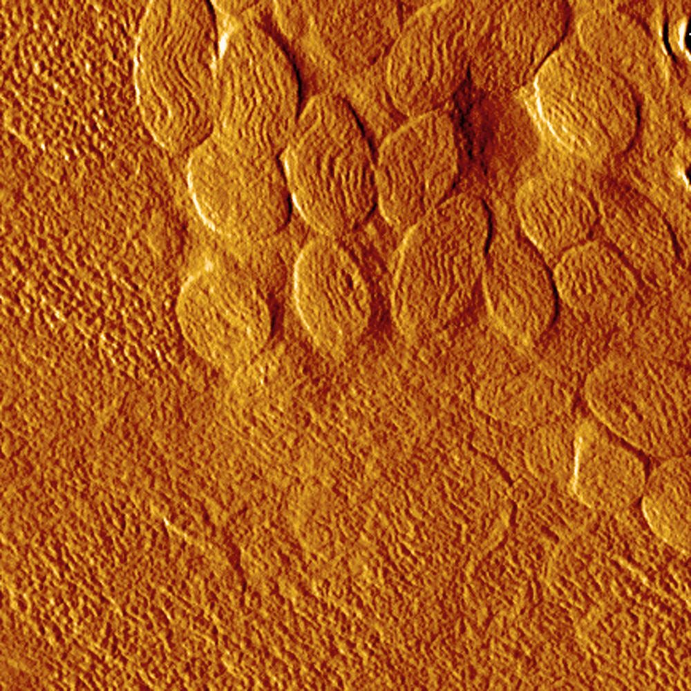
Morphology of a blend of two SBS block copolymers with different chain-architecture. AFM tapping mode, phase image, image size = 3 x 3 μm. Rameshwar Adhikari, Institut für Werkstoffwissenschaft, Martin-Luther-Universität, Halle-Wittenberg. Knife used: cryo AFM
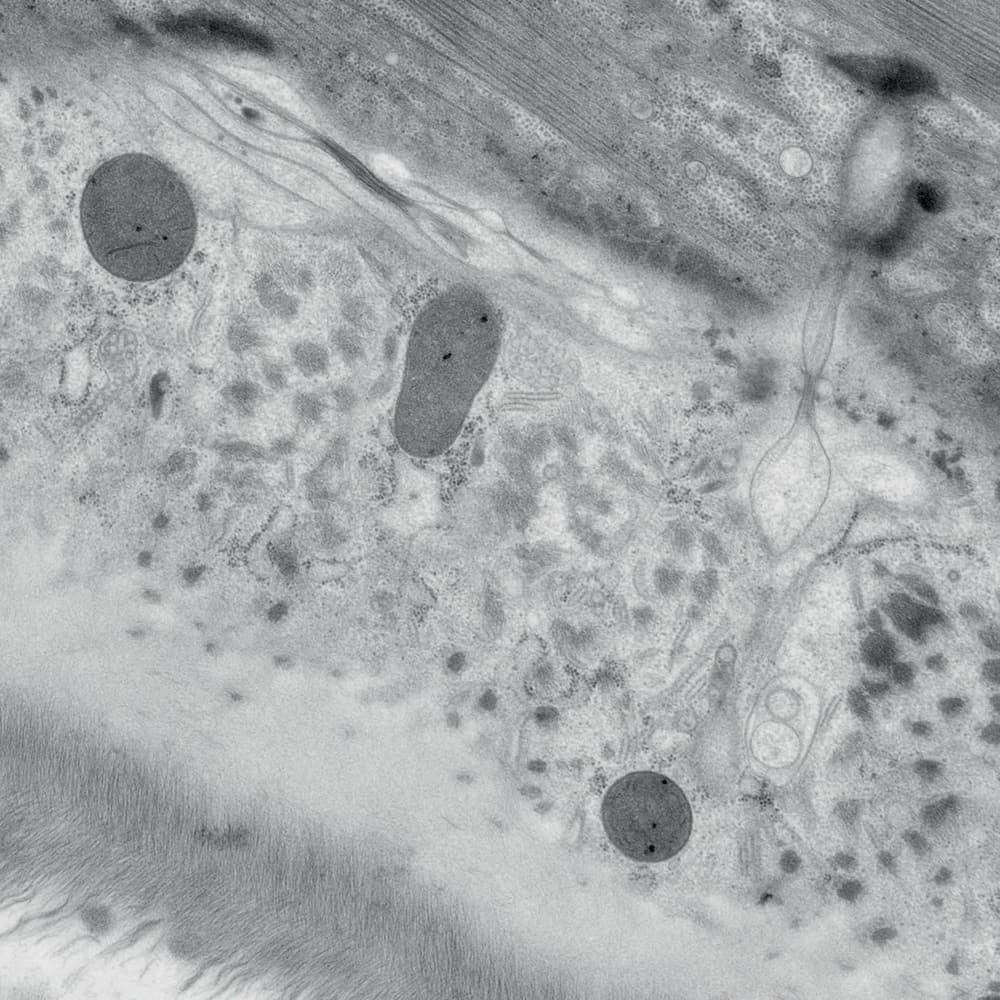
Ultrastructure of the roundworm Caenorhabditis elegans. Thomas Müller-Reichert, EM Technology Development, MPI Dresden, and Kent McDonald, Electron Microscopy Laboratory, University of California, Berkley. Scale bar: 28 mm = 1 μm. Knife used: ultra 45
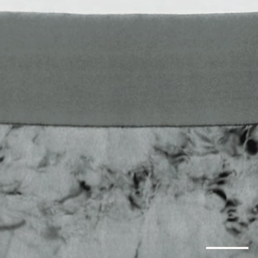
EM micrograph of an ultramicrotomed section of the anodic alumina fi lm formed on Al-2 wt%Cu alloy. Scale bar = 100 nm. Xiarong Zhou, School of Materials, University of Manchester. Knife used: ultra 35°
Polycarbonate modified with rubber Jens Sicking, Bayer Technology Services, Leverkusen. Knife used: ultra sonic
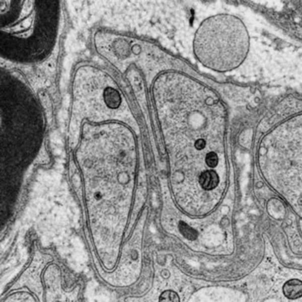
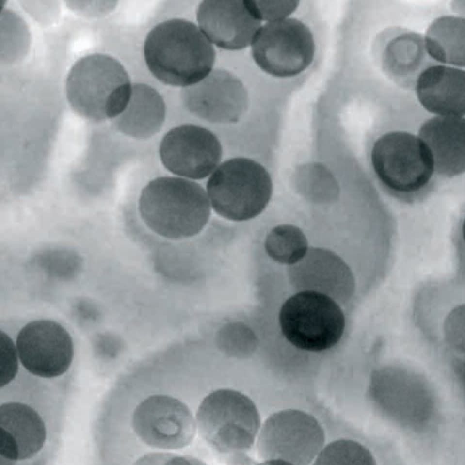
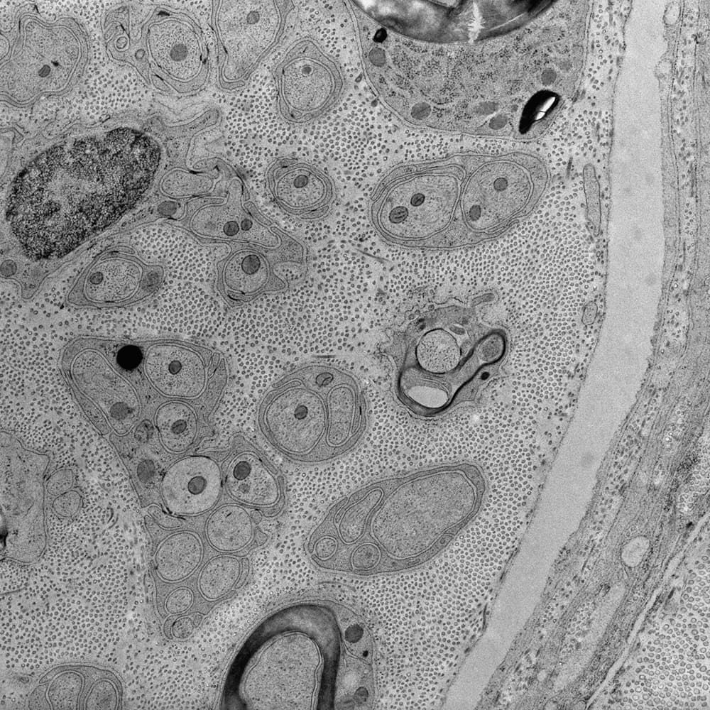
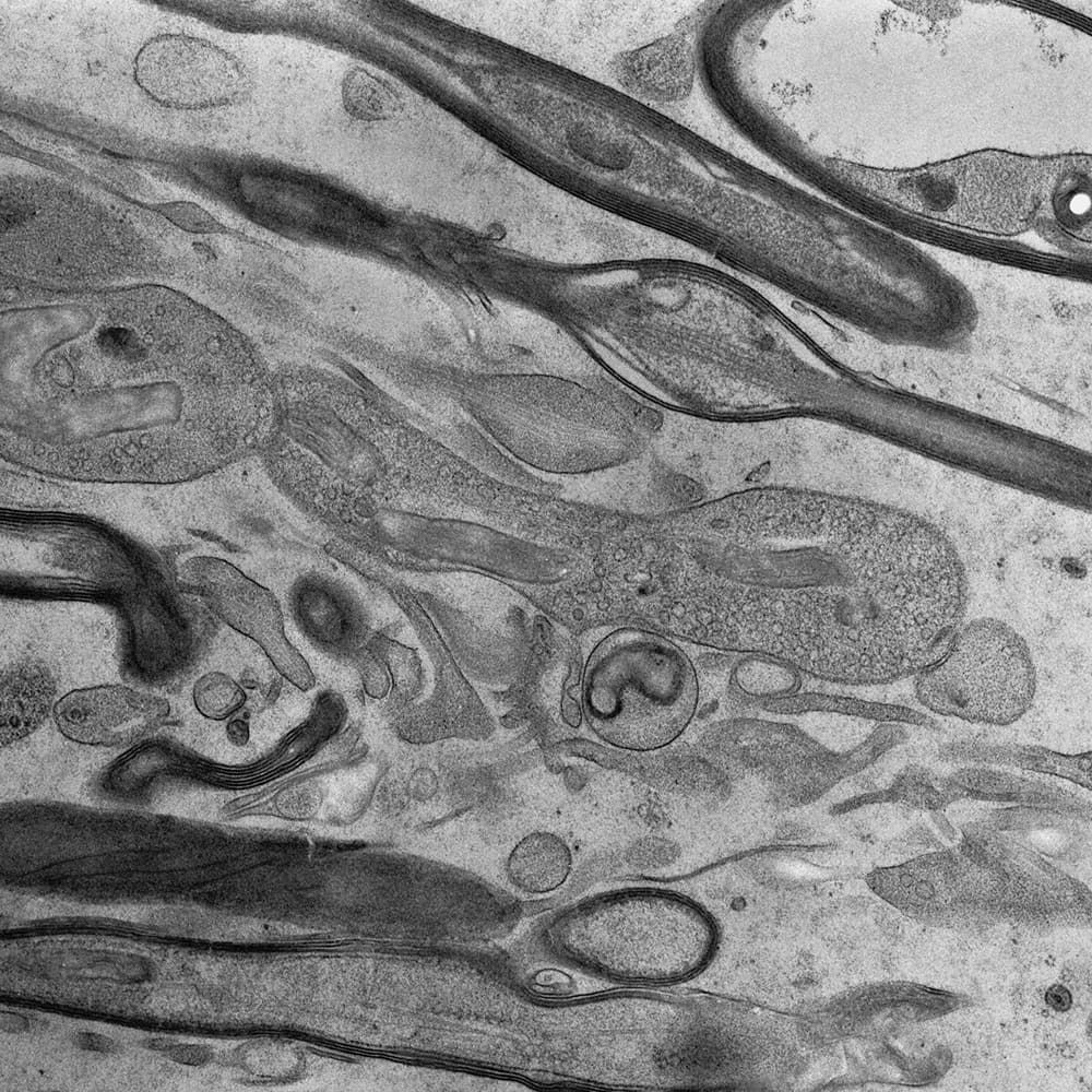
Peripheral nerve (rat), HP frozen, freeze substituted, Epon embedded, cut with the ultra sonic knife, section thickness 50nm. W. Graber, Institut of Anatomy, University of Bern, Switzerland. Knife used: ultra sonic
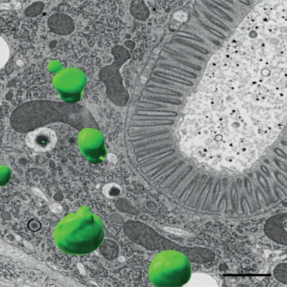
Section obtained with the ultra sonic Maxi knife. Scale bar 1 µm. Knife used: ultra sonic Maxi
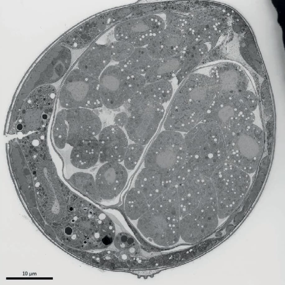
Serial section obtained with the ultra sonic Maxi knife at 55 nm from a high pressure frozen nematode sample. Knife used: ultra sonic Maxi
AFM amplitude image of the muscle of cat‘s mite Otodectes cynotis. The contrast covers amplitude variation in the 1 – 3 nm range. Size of the whole image equals 4.6 microns. Nadejda Borisovna Matsko, Institut für angewandte Physik, ETH Zürich. Knife used: ultra AFM
Knife used: ultra 45° Jumbo
Knife used: ultra 45° Jumbo


